Warning
You are using a web browser that we do not support. Our website will not work properly. Please update to a newer version or download a new web browser, such as Chrome or Firefox.
Help
Contact us
MyPDB
Sign in with Google
Sign in with Facebook
Sign in with ORCID
Help
Toggle navigation
RCSB PDB
Deposit
Prepare Data
PDBx/mmCIF file
pdb_extract
SF-Tool
Ligand Expo
MAXIT
Validate Data
Validation Server
Validation API
Information for Journals
Validation Task Forces
Deposit Data
wwPDB OneDep System
PDB-IHM
Help and Resources
Deposit FAQ
Validation FAQ
Tutorials
Annotation Policies
Processing Procedures
PDBx/mmCIF Dictionary
PDBx/mmCIF User Guide
Chemical Component Dictionary
Biologically Interesting Molecule Reference Dictionary (BIRD)
BioSync/Beamlines/Facilities
Related Tools
Search
Advanced Search
Sequence Similarity Search
Structure Similarity Search
Chemical Similarity Search
Chemical Sketch Tool
Browse by Annotations
New Entries
Unreleased Entries
PDB Statistics
Visualize
Mol* (MolStar)
Sequence Annotations Viewer
Genome View
Analyze
Pairwise Structure Alignment
Symmetry Resources in the PDB
Structure Quality
Grouping Structures
PDB Citation MeSH Network Explorer
PDB Statistics
EPPIC Biological Assemblies
External Data and Resources
Integrated Resources
Additional Resources
Download
Coordinates and Experimental Data
Sequences
Ligands
File Download Services
Web APIs
Learn
Training Courses
Guide to PDB Data
Molecule of the Month
Educational Resources
Curricula
Browse
News
SciArt Galleries
Irving Geis
David Goodsell
About
Contact Us
About RCSB PDB
Vision and Mission
Citation, Usage, Privacy Policies, Logo
News
PDB History
PDB50
User Community
Publications
RCSB PDB Advisory Committee
Team Members
Diversity, Equity, Inclusion, and Access
Service Status
Careers
COVID-19
234,440
Structures from the PDB
1,068,577
Computed Structure Models (CSM)
Include CSM
Advanced Search
|
Browse Annotations
Help
Search
Query History
Browse Annotations
MyPDB
QUERY:
PubMed ID = 30679383
MyPDB Login
Search API
Advanced Search Query Builder
Help
Full Text
Structure Attributes
Help
x
is any of
is not empty
NOT
Count
Add Subquery
Chemical Attributes
Sequence Similarity
Sequence Motif
Structure Similarity
Structure Motif
Chemical Similarity
Return
Structures
Polymer Entities
Assemblies
Non-polymer Entities
Molecular Definitions
grouped by
No Grouping
PDB Deposit Group ID
Include Computed Structure Models (CSM)
Count
Clear
Search
Search Summary
This query matches 10
Structures
.
Refinements
Structure Determination Methodology
experimental (10)
Scientific Name of Source Organism
Escherichia phage T7 (10)
Escherichia coli (1)
Taxonomy
Duplodnaviria (10)
Bacteria (1)
Experimental Method
ELECTRON MICROSCOPY (10)
Polymer Entity Type
DNA (10)
Protein (10)
RNA (4)
Refinement Resolution (Å)
3.0 - 3.5 (1)
3.5 - 4.0 (4)
4.0 - 4.5 (3)
> 4.5 (2)
Release Date
2015 - 2019 (10)
Enzyme Classification Name
Hydrolases (10)
Transferases (10)
Symmetry Type
Asymmetric (10)
-- Tabular Report --
Entry IDs
Create Custom Report
Structure
Sequence
Ligand
Oligosaccharide
Structural Genomics Center
Primary Citation
Biological Details
Sequence Clusters
Binding Affinity
Crystallization
Data Collection
Refinement
Refinement Parameters
Unit Cell
NMR Representative Model
NMR Spectrometer
NMR Sample Conditions
NMR Refinement
NMR Ensemble
NMR Software
EM Structure
All
Selected
1 to 10 of 10
Structures
Page 1 of 1
25
50
100
Sort by
↓ Score
↓ Release Date: Newest to Oldest
↑ Release Date: Oldest to Newest
↑ Entry ID: A to Z
↓ Entry ID: Z to A
↑ Resolution: Best to Worst
↓ Resolution: Worst to Best
↓ Priority: Experimental First
↑ Priority: Experimental Last
↓ Global pLDDT Score: Best to Worst
↑ Global pLDDT Score: Worst to Best
↑ Entry All Residues: Less to More
↓ Entry All Residues: More to Less
↑ Entry Modeled Residues: Less to More
↓ Entry Modeled Residues: More to Less
↑ Entry Chain Count: Less to More
↓ Entry Chain Count: More to Less
Explore in 3D
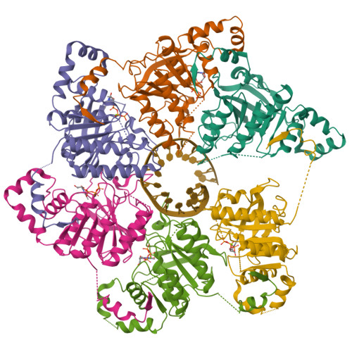
6N7I
|
pdb_00006n7i
Download File
View File
Structure of bacteriophage T7 E343Q mutant gp4 helicase-primase in complex with ssDNA, dTTP, AC dinucleotide and CTP (gp4(5)-DNA)
Gao, Y.
,
Cui, Y.
,
Zhou, Z.
,
Yang, W.
(2019) Science
363
:
Released
2019-03-06
Method
ELECTRON MICROSCOPY 3.2 Å
Organisms
Escherichia phage T7
Macromolecule
DNA primase/helicase
(protein)
DNA (25-MER)
(nucleic acid)
Unique Ligands
MG
,
TTP
Explore in 3D
6N7N
|
pdb_00006n7n
Download File
View File
Structure of bacteriophage T7 E343Q mutant gp4 helicase-primase in complex with ssDNA, dTTP, AC dinucleotide and CTP (form I)
Gao, Y.
,
Cui, Y.
,
Zhou, Z.
,
Yang, W.
(2019) Science
363
:
Released
2019-03-06
Method
ELECTRON MICROSCOPY 3.5 Å
Organisms
Escherichia phage T7
Macromolecule
DNA primase/helicase
(protein)
DNA (5'-D(P*TP*TP*TP*TP*TP*TP*TP*TP*TP*TP*TP*TP*TP*TP*T)-3')
(nucleic acid)
Unique Ligands
MG
,
TTP
Explore in 3D
6N7S
|
pdb_00006n7s
Download File
View File
Structure of bacteriophage T7 E343Q mutant gp4 helicase-primase in complex with ssDNA, dTTP, AC dinucleotide and CTP (form II)
Gao, Y.
,
Cui, Y.
,
Zhou, Z.
,
Yang, W.
(2019) Science
363
:
Released
2019-03-06
Method
ELECTRON MICROSCOPY 4.6 Å
Organisms
Escherichia phage T7
Macromolecule
DNA primase/helicase
(protein)
DNA (25-MER)
(nucleic acid)
Unique Ligands
MG
,
TTP
Explore in 3D
6N7T
|
pdb_00006n7t
Download File
View File
Structure of bacteriophage T7 E343Q mutant gp4 helicase-primase in complex with ssDNA, dTTP, AC dinucleotide and CTP (form III)
Gao, Y.
,
Cui, Y.
,
Zhou, Z.
,
Yang, W.
(2019) Science
363
:
Released
2019-03-06
Method
ELECTRON MICROSCOPY 3.9 Å
Organisms
Escherichia phage T7
Macromolecule
DNA primase/helicase
(protein)
DNA (25-MER)
(nucleic acid)
Unique Ligands
MG
,
TTP
Explore in 3D
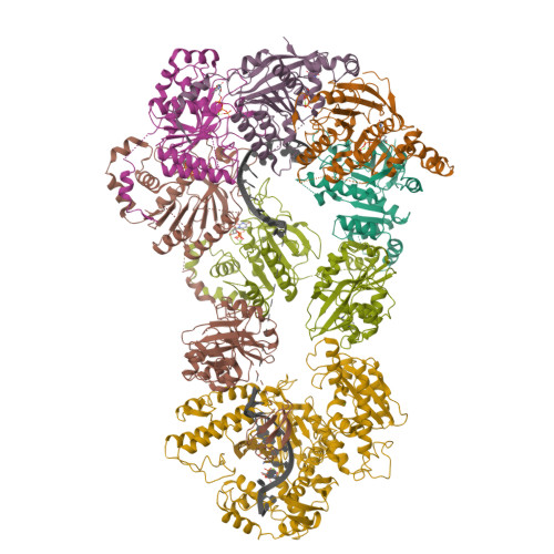
6N7V
|
pdb_00006n7v
Download File
View File
Structure of bacteriophage T7 gp4 (helicase-primase, E343Q mutant) in complex with ssDNA, dTTP, AC dinucleotide, and CTP (from multiple lead complexes)
Gao, Y.
,
Fox, T.
,
Val, N.
,
Yang, W.
(2019) Science
363
:
Released
2019-03-06
Method
ELECTRON MICROSCOPY 3.8 Å
Organisms
Escherichia phage T7
Macromolecule
DNA primase/helicase
(protein)
DNA (93-MER)
(nucleic acid)
Unique Ligands
MG
,
TTP
Explore in 3D
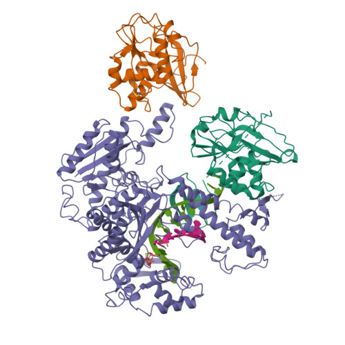
6N7W
|
pdb_00006n7w
Download File
View File
Structure of bacteriophage T7 leading-strand DNA polymerase (D5A/E7A)/Trx in complex with a DNA fork and incoming dTTP (from multiple lead complexes)
Gao, Y.
,
Fox, T.
,
Val, N.
,
Yang, W.
(2019) Science
363
:
Released
2019-03-06
Method
ELECTRON MICROSCOPY 4.5 Å
Organisms
Escherichia coli
Escherichia phage T7
Macromolecule
DNA-directed DNA polymerase
(protein)
TrxA
(protein)
DNA (25-MER)
(nucleic acid)
DNA (77-MER)
(nucleic acid)
Unique Ligands
MG
,
TTP
Explore in 3D
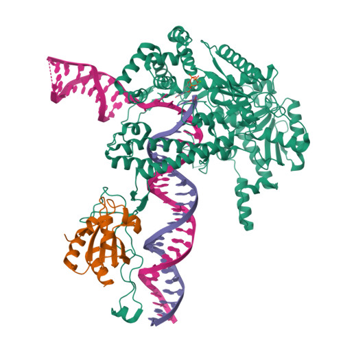
6N9U
|
pdb_00006n9u
Download File
View File
Structure of bacteriophage T7 lagging-strand DNA polymerase (D5A/E7A) interacting with primase domains of two gp4 subunits bound to an RNA/DNA hybrid and dTTP (from LagS1)
Gao, Y.
,
Fox, T.
,
Val, N.
,
Yang, W.
(2019) Science
363
:
Released
2019-03-06
Method
ELECTRON MICROSCOPY 3.7 Å
Organisms
Escherichia phage T7
Macromolecule
DNA primase/helicase
(protein)
DNA-directed DNA polymerase
(protein)
DNA (44-MER)
(nucleic acid)
RNA (5'-R(*AP*CP*CP*AP*G)-D(P*(DOC))-3')
(nucleic acid)
Unique Ligands
MG
,
TTP
,
ZN
Explore in 3D
6N9V
|
pdb_00006n9v
Download File
View File
Structure of bacteriophage T7 lagging-strand DNA polymerase (D5A/E7A) and gp4 (helicase/primase) bound to DNA including RNA/DNA hybrid, and an incoming dTTP (LagS1)
Gao, Y.
,
Fox, T.
,
Val, N.
,
Yang, W.
(2019) Science
363
:
Released
2019-03-06
Method
ELECTRON MICROSCOPY 4 Å
Organisms
Escherichia phage T7
Macromolecule
DNA primase/helicase
(protein)
DNA-directed DNA polymerase
(protein)
Primer
(nucleic acid)
Template
(nucleic acid)
Unique Ligands
MG
,
TTP
,
ZN
Explore in 3D
6N9W
|
pdb_00006n9w
Download File
View File
Structure of bacteriophage T7 lagging-strand DNA polymerase (D5A/E7A) and gp4 (helicase/primase) bound to DNA including RNA/DNA hybrid, and an incoming dTTP (LagS2)
Gao, Y.
,
Cui, Y.
,
Zhou, Z.
,
Yang, W.
(2019) Science
363
:
Released
2019-03-06
Method
ELECTRON MICROSCOPY 4 Å
Organisms
Escherichia phage T7
Macromolecule
DNA primase/helicase
(protein)
DNA-directed DNA polymerase
(protein)
Primer
(nucleic acid)
Template
(nucleic acid)
Unique Ligands
MG
,
TTP
,
ZN
Explore in 3D
6N9X
|
pdb_00006n9x
Download File
View File
Structure of bacteriophage T7 lagging-strand DNA polymerase (D5A/E7A) and gp4 (helicase/primase) bound to DNA including RNA/DNA hybrid, and an incoming dTTP (LagS3)
Gao, Y.
,
Fox, T.
,
Val, N.
,
Yang, W.
(2019) Science
363
:
Released
2019-03-06
Method
ELECTRON MICROSCOPY 4.1 Å
Organisms
Escherichia phage T7
Macromolecule
DNA primase/helicase
(protein)
DNA-directed DNA polymerase
(protein)
Primer
(nucleic acid)
Template
(nucleic acid)
Unique Ligands
MG
,
TTP
,
ZN
1 to 10 of 10
Structures
Page 1 of 1
25
50
100
Sort by
↓ Score
↓ Release Date: Newest to Oldest
↑ Release Date: Oldest to Newest
↑ Entry ID: A to Z
↓ Entry ID: Z to A
↑ Resolution: Best to Worst
↓ Resolution: Worst to Best
↓ Priority: Experimental First
↑ Priority: Experimental Last
↓ Global pLDDT Score: Best to Worst
↑ Global pLDDT Score: Worst to Best
↑ Entry All Residues: Less to More
↓ Entry All Residues: More to Less
↑ Entry Modeled Residues: Less to More
↓ Entry Modeled Residues: More to Less
↑ Entry Chain Count: Less to More
↓ Entry Chain Count: More to Less
About
About Us
Citing Us
Publications
Team
Careers
Usage & Privacy
Support
Contact Us
Help
Website FAQ
Glossary
Service Status
RCSB PDB is hosted by
RCSB PDB is a member of the
RCSB Partners
Nucleic Acid Knowledgebase
wwPDB Partners
RCSB PDB
PDBe
PDBj
BMRB
EMDB