Warning You are using a web browser that we do not support. Our website will not work properly. Please update to a newer version or download a new web browser, such as Chrome or Firefox.
Help
Contact us
  • Sign in with Google
  • Sign in with Facebook
  • Sign in with ORCID

  • Help
RCSB PDB
  • Deposit 

      Prepare Data

    • PDBx/mmCIF file
    • pdb_extract
    • SF-Tool
    • Ligand Expo
    • MAXIT

      Validate Data

    • Validation Server
    • Validation API
    • Information for Journals
    • Validation Task Forces

      Deposit Data

    • wwPDB OneDep System
    • PDB-IHM

      Help and Resources

    • Deposit FAQ
    • Validation FAQ
    • Tutorials
    • Annotation Policies
    • Processing Procedures
    • PDBx/mmCIF Dictionary
    • PDBx/mmCIF User Guide
    • Chemical Component Dictionary
    • Biologically Interesting Molecule Reference Dictionary (BIRD)
    • BioSync/Beamlines/Facilities
    • Related Tools
  • Search 
    • Advanced Search
    • Sequence Similarity Search
    • Structure Similarity Search
    • Chemical Similarity Search
    • Chemical Sketch Tool
    • Browse by Annotations
    • New Entries
    • Unreleased Entries
    • PDB Statistics
  • Visualize 
    • Mol* (MolStar)
    • Sequence Annotations Viewer
    • Genome View
  • Analyze 
    • Pairwise Structure Alignment
    • Symmetry Resources in the PDB
    • Structure Quality
    • Grouping Structures
    • PDB Citation MeSH Network Explorer
    • PDB Statistics
    • EPPIC Biological Assemblies
    • External Data and Resources
    • Integrated Resources
    • Additional Resources
  • Download 
    • Coordinates and Experimental Data
    • Sequences
    • Ligands
    • File Download Services
    • Web APIs
  • Learn 

    • Training Courses
    • Guide to PDB Data
    • Molecule of the Month
    • Educational Resources
    • Curricula
    • Browse
    • News
    • SciArt Galleries
    • Irving Geis
    • David Goodsell
  • About 
    • Contact Us
    • About RCSB PDB
    • Vision and Mission
    • Citation, Usage, Privacy Policies, Logo
    • News
    • PDB History
    • PDB50
    • User Community
    • Publications
    • RCSB PDB Advisory Committee
    • Team Members
    • Service Status
  • Careers
  • COVID-19
RCSB PDB
235,666 Structures from the PDB
1,068,577 Computed Structure Models (CSM)
Include CSM
Advanced Search
|
Browse Annotations
Help
  • PDB-101
  • wwPDB
  • EMDataResource
  • NAKB: Nucleic Acid Knowledgebase
  • wwPDB Foundation
  • PDB-IHM Logo
  • Search
  • Query History
  • Browse Annotations
  • MyPDB
QUERY: BIRD Name HAS EXACT PHRASE "=gamma-cyclodextrin"
MyPDB Login
 Search API
Advanced Search Query Builder
Help
Full Text
Structure Attributes
Chemical Attributes
Help
x
NOT
Count
Add Subquery
Sequence Similarity
Sequence Motif
Structure Similarity
Structure Motif
Chemical Similarity
Return
grouped by
Include Computed Structure Models (CSM)
Count
Clear
Search
Search Summary This query matches 8 Structures.
Refinements
Structure Determination Methodology
Scientific Name of Source Organism
Taxonomy
Experimental Method
Polymer Entity Type
Refinement Resolution (Å)
Release Date
Enzyme Classification Name
Symmetry Type
SCOP Classification
1 to 8 of 8 Structures
Page 1 of 1
Sort by
Gallery item
 Explore in 3D

1D3C | pdb_00001d3c

Download FileView File

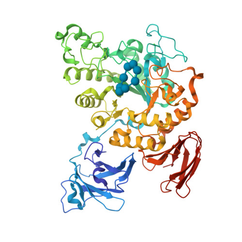
MICHAELIS COMPLEX OF BACILLUS CIRCULANS STRAIN 251 CYCLODEXTRIN GLYCOSYLTRANSFERASE WITH GAMMA-CYCLODEXTRIN

Uitdehaag, J.C.M., Kalk, K.H., van der Veen, B.A., Dijkhuizen, L., Dijkstra, B.W.

(1999) J Biological Chem 274: 34868-34876

Released1999-12-22
Method
X-RAY DIFFRACTION 1.78 Å
Organisms
Niallia circulans
Macromolecule
CYCLODEXTRIN GLYCOSYLTRANSFERASE (protein)
Unique LigandsCA, MPD
Unique branched monosaccharidesGLC
Gallery item
 Explore in 3D

1P2G | pdb_00001p2g

Download FileView File

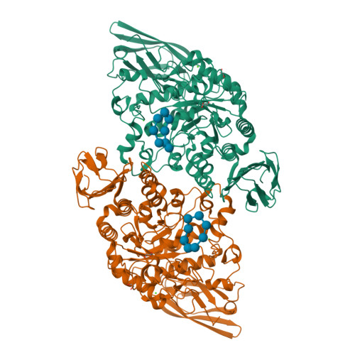
Crystal Structure of Glycogen Phosphorylase B in complex with Gamma Cyclodextrin

Pinotsis, N., Leonidas, D.D., Chrysina, E.D., Oikonomakos, N.G., Mavridis, I.M.

(2003) Protein Sci 12: 1914-1924

Released2003-09-02
Method
X-RAY DIFFRACTION 2.3 Å
Organisms
Oryctolagus cuniculus
Macromolecule
Glycogen phosphorylase, muscle form (protein)
Unique LigandsPLP
Unique branched monosaccharidesGLC
Gallery item
 Explore in 3D

1VFU | pdb_00001vfu

Download FileView File

Crystal structure of Thermoactinomyces vulgaris R-47 amylase 2/gamma-cyclodextrin complex

Ohtaki, A., Mizuno, M., Tonozuka, T., Sakano, Y., Kamitori, S.

(2004) J Biological Chem 279: 31033-31040

Released2005-02-08
Method
X-RAY DIFFRACTION 3.1 Å
Organisms
Thermoactinomyces vulgaris
Macromolecule
Neopullulanase 2 (protein)
Unique LigandsCA
Unique branched monosaccharidesGLC
Gallery item
 Explore in 3D

2ZYK | pdb_00002zyk

Download FileView File

Crystal structure of cyclo/maltodextrin-binding protein complexed with gamma-cyclodextrin

Tonozuka, T., Sogawa, A., Yamada, M., Matsumoto, N., Yoshida, H., Kamitori, S., Ichikawa, K., Mizuno, M., Nishikawa, A., Sakano, Y.

(2007) FEBS J 274: 2109-2120

Released2009-02-10
Method
X-RAY DIFFRACTION 2.5 Å
Organisms
Thermoactinomyces vulgaris
Macromolecule
Solute-binding protein (protein)
Unique branched monosaccharidesGLC
Gallery item
 Explore in 3D

3EDK | pdb_00003edk

Download FileView File

Structural base for cyclodextrin hydrolysis

Buedenbender, S., Schulz, G.E.

(2009) J Mol Biology 385: 606-617

Released2009-03-03
Method
X-RAY DIFFRACTION 1.77 Å
Organisms
Flavobacterium sp. 92
Macromolecule
Cyclomaltodextrinase (protein)
Unique LigandsCA, GOL
Unique branched monosaccharidesGLC
Gallery item
 Explore in 3D

5E70 | pdb_00005e70

Download FileView File

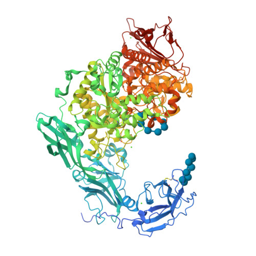
Crystal structure of Ecoli Branching Enzyme with gamma cyclodextrin

Feng, L., Nosrati, M., Geiger, J.H.

(2016) Acta Crystallogr D Struct Biol 72: 641-647

Released2015-12-16
Method
X-RAY DIFFRACTION 2.33 Å
Organisms
Escherichia coli O139:H28 str. E24377A
Macromolecule
1,4-alpha-glucan branching enzyme GlgB (protein)
Unique LigandsGOL
Unique branched monosaccharidesGLC
Gallery item
 Explore in 3D

5MKA | pdb_00005mka

Download FileView File

Maltodextrin binding protein MalE1 from L. casei BL23 bound to gamma-cyclodextrin

Homburg, C., Bommer, M., Wuttge, S., Hobe, C., Beck, S., Dobbek, H., Deutscher, J., Licht, A., Schneider, E.

(2017) Mol Microbiol 105: 25-45

Released2017-07-19
Method
X-RAY DIFFRACTION 1.149 Å
Organisms
Lacticaseibacillus casei
Macromolecule
MalE1 (protein)
Unique branched monosaccharidesGLC
Gallery item
 Explore in 3D

5YNH | pdb_00005ynh

Download FileView File

Crystal structure of Pullulanase from Klebsiella pneumoniae complex at 10 mM gamma-cyclodextrin

Saka, N., Iwamoto, H., Takahashi, N., Mizutani, K., Mikami, B.

(2018) Acta Crystallogr D Struct Biol 74: 1115-1123

Released2018-10-24
Method
X-RAY DIFFRACTION 2.05 Å
Organisms
Klebsiella pneumoniae
Macromolecule
PulA protein (protein)
Unique LigandsCA, GOL
Unique branched monosaccharidesGLC
1 to 8 of 8 Structures
Page 1 of 1
Sort by
  • About
  • About Us
  • Citing Us
  • Publications
  • Team
  • Careers
  • Usage & Privacy
  • Support
  • Contact Us
  • Help
  • Website FAQ
  • Glossary
  • Service Status
  • RCSB PDB is hosted by

  • The Rutgers Artificial Intelligence and Data Science Collaboratory logo
    Uiversity of California San Diego logoSan Diego Supercomputer Center logo
    University of California San Francisco Logo
  • RCSB PDB is a member of the

  • wwPDB
    EMDataResource

  • RCSB Partners
  • Nucleic Acid Knowledgebase
  • wwPDB Partners
  • RCSB PDB
  • PDBe
  • PDBj
  • BMRB
  • EMDB
CoreTrustSeal
Global Biodata Coalition Logo

RCSB PDB Core Operations are funded by the U.S. National Science Foundation (DBI-2321666), the US Department of Energy (DE-SC0019749), and the National Cancer Institute, National Institute of Allergy and Infectious Diseases, and National Institute of General Medical Sciences of the National Institutes of Health under grant R01GM157729.